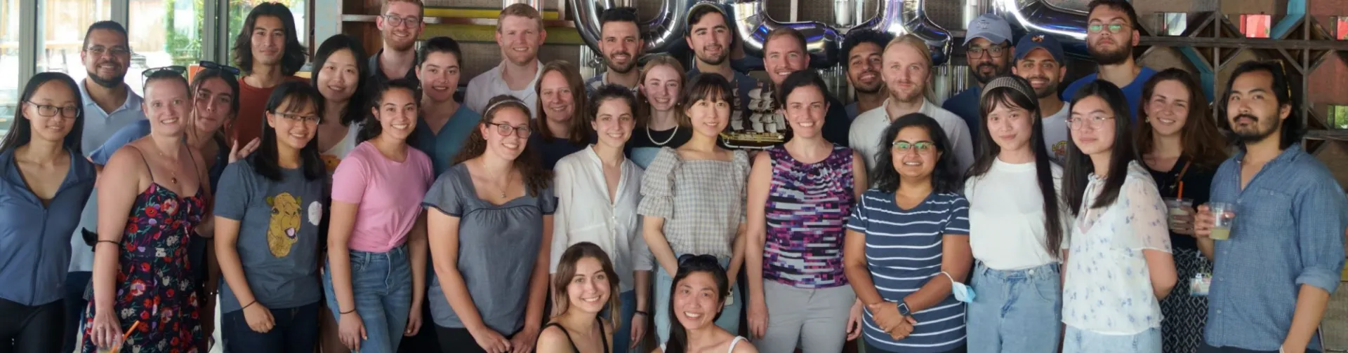
Welcome to the UEIL
Led by Prof. Elisa Konofagou, the Ultrasound and Elasticity Imaging Laboratory works on developing novel, ultrasound-based techniques for both imaging and therapeutic applications. Some of the ongoing research in the UEIL includes:
-
Enabling targeted drug delivery to the brain by opening the blood-brain barrier with focused ultrasound.
-
Mapping the mechanical and electromechanical properties of the heart using myocardial elastography and electromechanical wave imaging.
-
Monitoring High-Intensity Focused Ultrasound (HIFU) ablation therapy with harmonic motion imaging.
-
Using pulse wave imaging to quantify arterial wall mechanics and blood flow dynamics to characterize vascular diseases, including carotid atherosclerosis and hypertension.
-
Modulating neural activity in the central and peripheral nervous system through high-intensity focused ultrasound.
The UEIL is located in the Columbia University Medical Center at the New York Presbyterian Hospital and is part of the Department of Biomedical Engineering at Columbia University. If you have any additional inquiries, or if you would be interested in collaborating with our laboratory, please feel free to contact us.
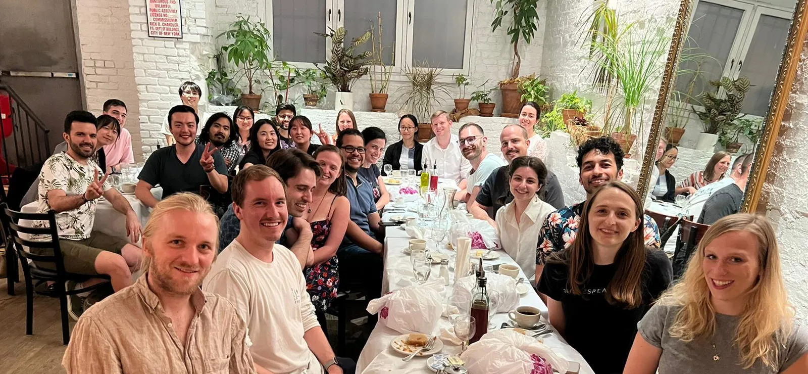
Latest Lab Events and Celebrations
Image Carousel with 12 slides
A carousel is a rotating set of images. Use the previous and next buttons to change the displayed slide
-
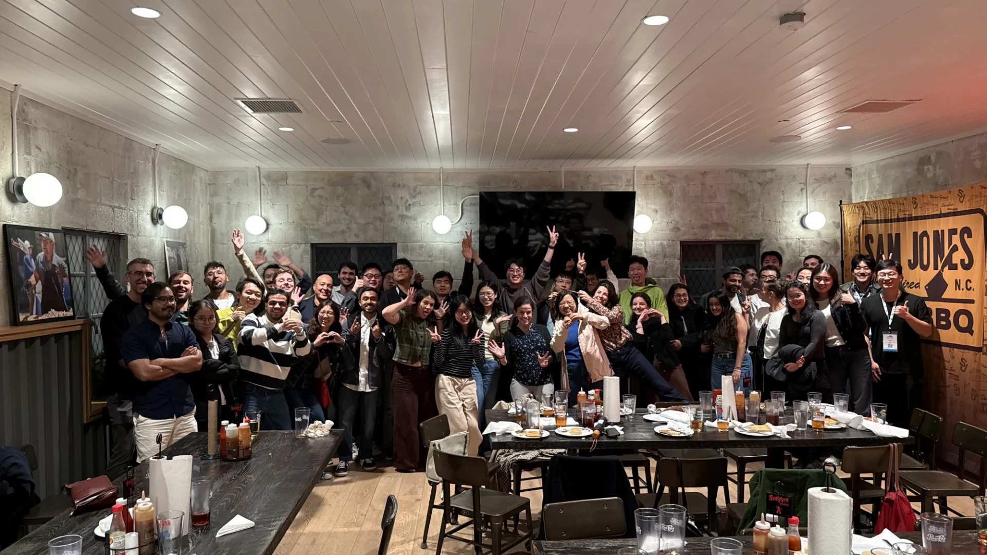
Slide 1: UEIL Reunion with Alumni at IUS 2026
-
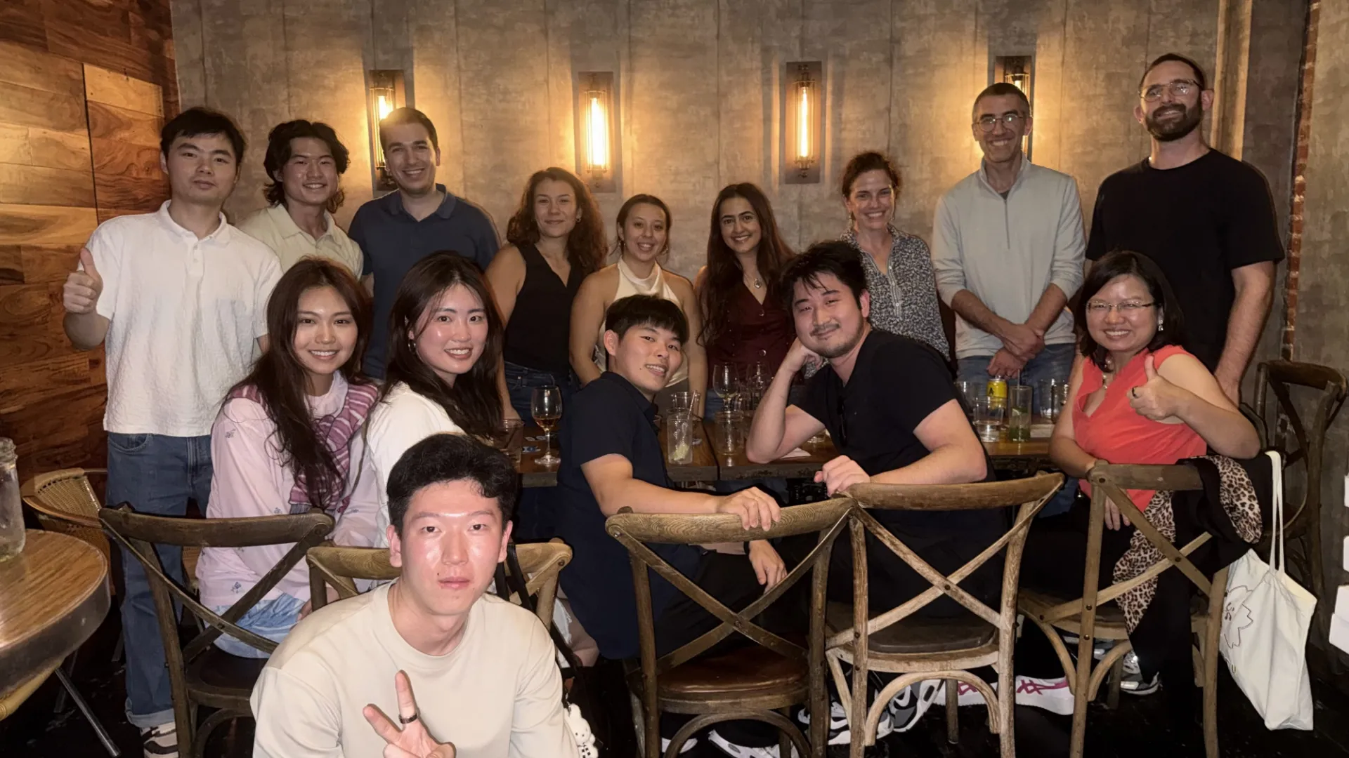
Slide 2: UEIL Members with Alumni
-
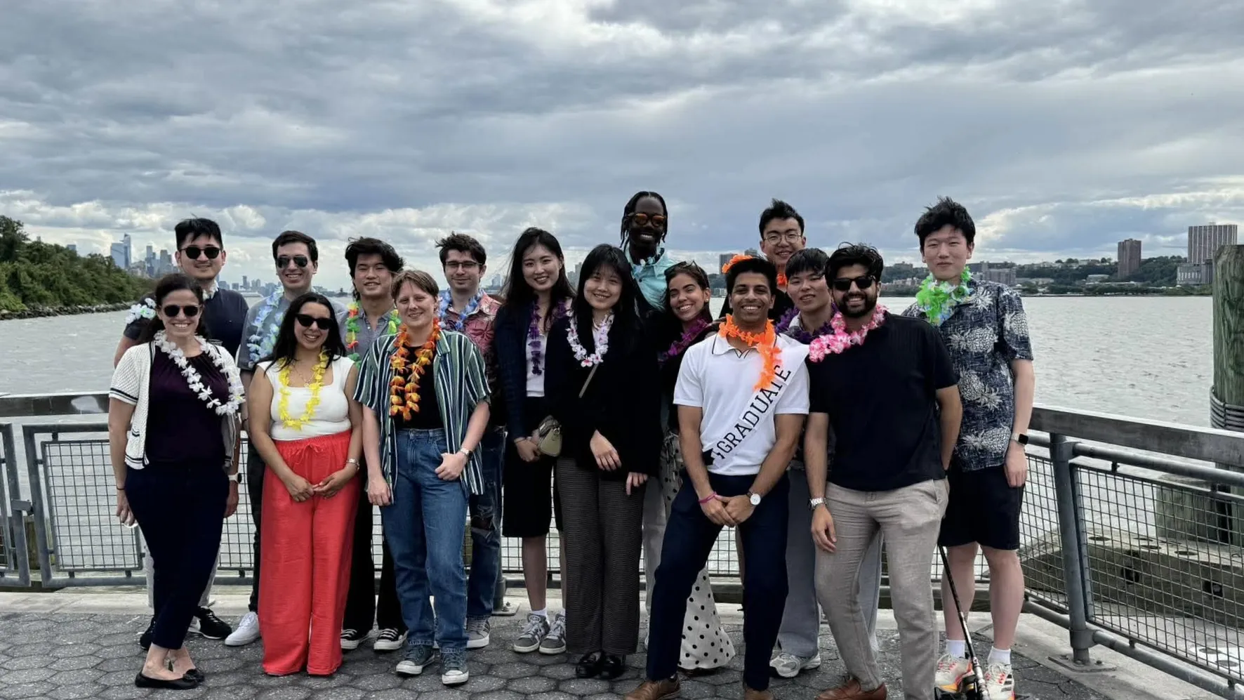
Slide 3: UEIL at the BME Department Celebration 2026
-
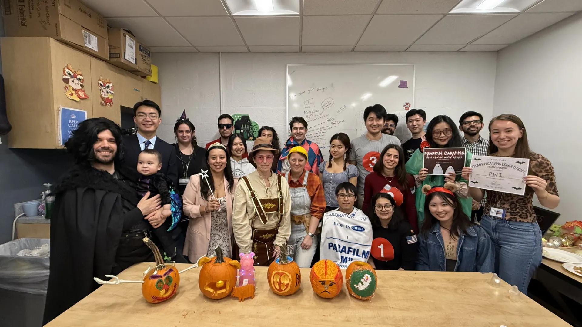
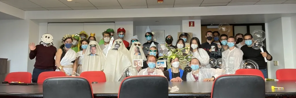
Slide 4: Lab Halloween 2025!
-
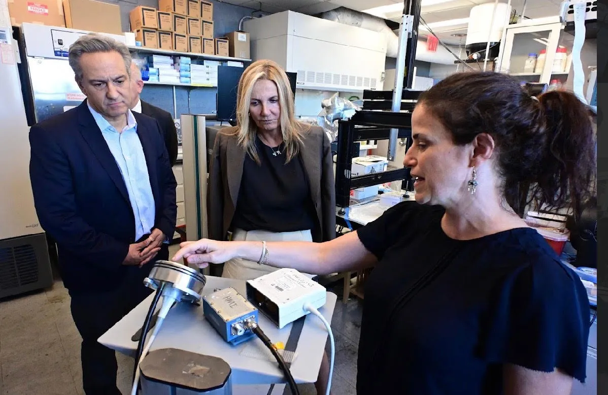
Slide 5: First Lady of Greece visits UEIL
-
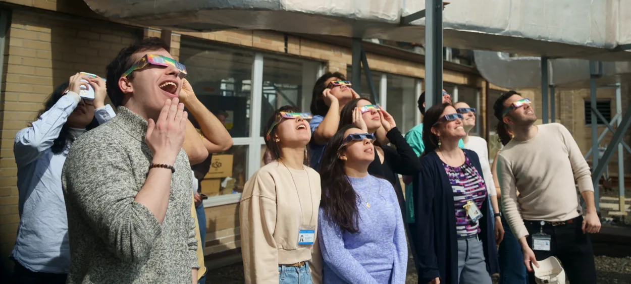
Slide 6: Solar Eclipse 2024!
-
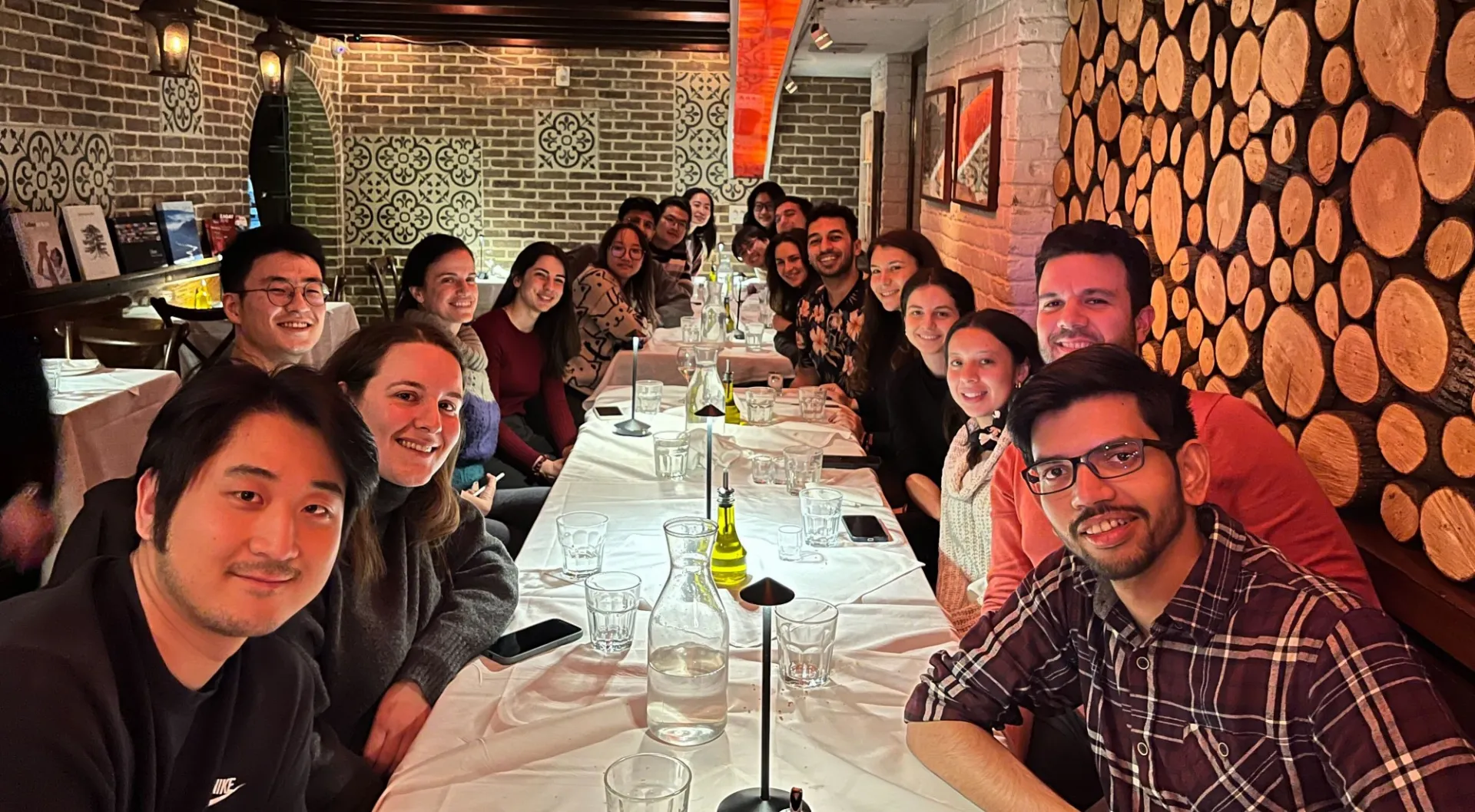
Slide 7: Jad's Defense Celebration Dinner! Congratulations Jad!
-
Slide 8: Recent and Soon-to-be Alumni Farewell
-
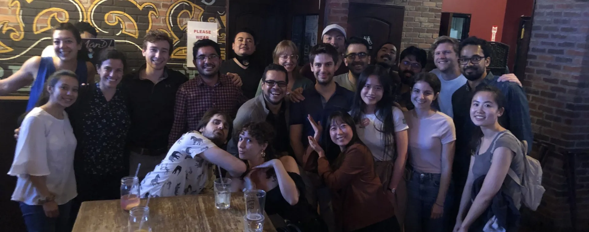
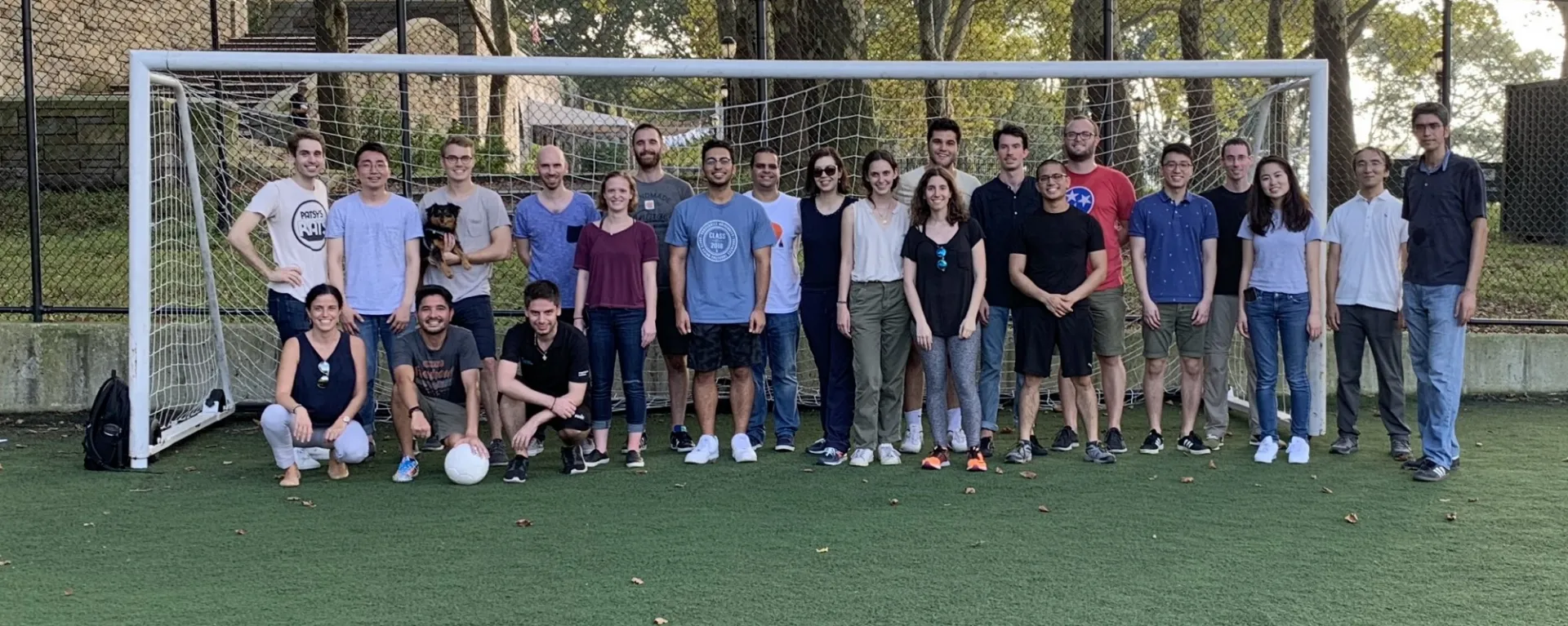
Slide 9: Annual Lab Outing
-
Slide 10: Halloween costume contest
-
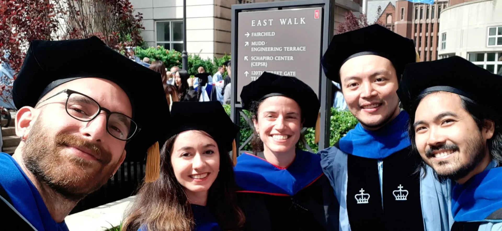
Slide 11: 2022 Commencement
-
Slide 12: Grigorios phd defense celebration! Congrats doc!
Latest News
UEIL Alumni Reunions in NYC and North Carolina!
A Recap of UEIL at IUS 2026
UEIL attended IUS 2026 with many posters and lectures! We had a lot of run in Raleigh, learned a lot during the conference, and cannot wait for next year's IUS!
UEIL is Attending IUS 2026!
The 2026 IEEE International Ultrasonics Symposium is held from Oct 4-8th in Raleigh, North Carolina, USA! UEIL is very excited to attend the conference - find details of their presentations and posters below: